The median umbilical ligament runs down the lower portion of the front of the abdominal wall. An umbilical cord is a thick blood-rich cord that connects a baby to its mother during the gestation process.
![]()
Medial Umbilical Ligament Anatomy Branches Supply Kenhub
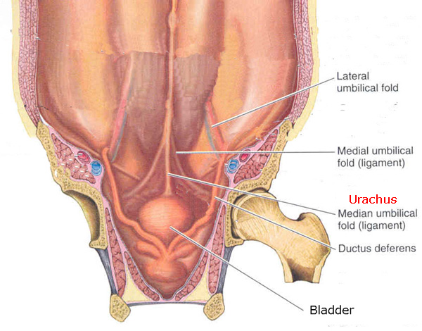
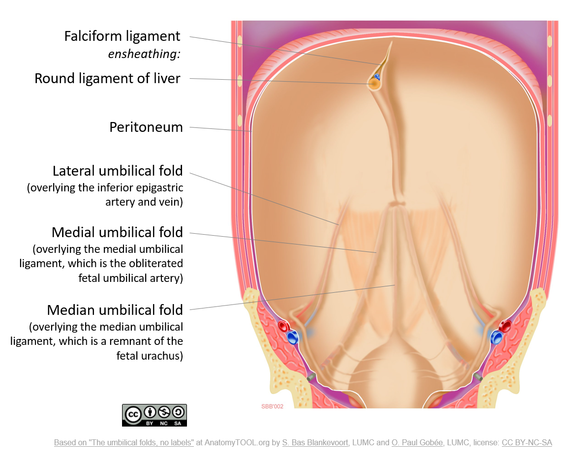
The median umbilical fold runs superiorly from the apex of the bladder to the umbilicus.

. It is a shrivelled piece of tissue that represents the remnant of the embryonic urachus. The median umbilical ligament is a structure in human anatomy. The median umbilical ligament or Xanders ligament is a structure in human anatomy.
These ligaments are all that remain of the umbilical cord. The paired medial umbilical folds pass from the pelvis to the umbilicus and cover the underlying medial umbilical ligaments. These structures are vestigial and for the most part do not serve a specific function in the body.
It extends from the apex of the bladder to the umbilicus on the deep surface of the anterior abdominal wall. The remnants of an embryonic communication between the allantois and cloaca. Just so what are the medial umbilical ligaments remnants of.
It is important to distinguish between the medial vs median umbilical ligaments. This fold is formed by the underlying median umbilical ligament. The medial umbilical ligament is an anatomic structure present in the human body that exists as a remnant of blood vessels that were important to fetal circulation.
The medial umbilical ligament is a paired structure found in human anatomy. Mean age was 821 years ranging between 56 and 96 years with a male-to-female ratio of 141. The median and medial umbilical ligaments form a peritoneal depression on each side of the urinary bladder referred to as the supravesical fossae.
Fortunately the anatomy itself is less confusing than the terminology. This area usually contains the fundus of the distended urinary bladder and can be clinically significant owing to the fact that the supravesical hernias can arise here. The median and medial umbilical ligaments were classified into four types based on their interrelationships.
It is different to the median umbilical ligament a structure that represents the remnant of. Inguinal swelling due to rare external supravesical hernia--a case report. The medial umbilical ligamentor cord of umbilical artery or obliterated umbilical artery is a paired structure found in human anatomy.
Lateral to this structure are the medial umbilical ligament and the lateral umbilical ligament. It then becomes the urachus in the fetus. Ninety-two per cent was white and 8 per cent black adults.
The urachus median umbilical ligament is visible along the anterior parietal peritoneum. It is on the deep surface of the anterior abdominal wall and is covered by the medial umbilical foldsplicae umbilicales mediales. It is also known as the cord of the umbilical artery.
As medial umbilical ligaments can develop chronic infection in the event of omphalitis the medial umbilical ligaments were resected continuously. It extends from the apex of the bladder to the umbilicus on the deep surface of the anterior abdominal wall. It is also formed from the cloaca in utero.
The median umbilical ligament has. There are also check ligaments restricting the vertical movements but the expansions of these muscles are thinner and less distinct than those of the horizontal recti muscles. Three different umbilical ligaments the median medial and lateral can be found in the abdominal anatomy legacies of the umbilical cord.
The medial umbilical ligaments are anatomical remnants of the obliterated foetal umbilical arteries. The most common type was the median umbilical ligament terminated by joining one or both medial umbilical ligaments Type II 411. The supravesical fossa is the area of abdominal wall between remnant of urachus Median umbilical ligament and remnant of left or right umbilical artery medial umbilical ligament.
Median medial lateral. After the cephalic side of the urachus and the medial umbilical ligaments were dropped into the abdominal cavity an intraperitoneal wound retractor Lap Protector Hakko Medical Nagano Japan. It consists of a cord often accompanied by vessels that must be controlled when dividing the urachus.
The urachus connects the dome of the bladder to the umbilical cord during fetal life and is located behind the abdominal wall and anterior to the peritoneum in the space of Retzius. The medial umbilical ligament is the aforementioned paired structure related to the umbilical arteries while the median umbilical ligament contains the urachus. Click to see full answer.
The medial rectus is attached to the lacrimal bone medial check ligament and the lateral rectus to the zygomatic bone lateral check ligament. A connecting branch arrow is evident between the LTH and the MdU that is on the left side. On either side laterally the median umbilical ligament is separated from the medial umbilical ligament by a peritoneal recess.
The folds are 2 of the 5 umbilical folds and should not be confused with the single midline median umbilical fold. The midline fold is called the median umbilical ligament and the paired paramedian folds are called the medial umbilical ligaments. Just so what becomes the median umbilical.
It is a remnant of the fetal urachus. Paired medial umbilical ligaments run along other side with a matching set of lateral ligaments. The median umbilical ligament begins as the allantois in the embryonic period.
The median umbilical ligament MnU and medial umbilical ligament MdU fuse into a band that continues to the ligamentum teres hepatis LTH covering the UR. In relation to the bladder the median umbilical fold is the most important of these structures as it contains the urachal ligament. It is on the deep surface of the anterior abdominal wall and is covered by the medial umbilical folds.
By birth the urachus is obliterated and becomes a vestigial structure known as. The bilateral supravesical fossae lie between the median and bilateral medial umbilical folds. The medial umbilical ligament or cord of umbilical artery or obliterated umbilical artery is a paired structure found in human anatomy.
It is different from the median umbilical ligament a structure that represents the. It is on the deep surface of the anterior abdominal wall and is covered by the medial umbilical folds plicae umbilicales mediales. This later develops into the median umbilical ligament at birth.
Round hepatic median and medial ligaments umbilical ring umbilical and umbilicovesicular fasciae and pattern of attachment to the ring were dissected and measured. It is a shrivelled piece of tissue that represents the remnant of the embryonic urachus.
Epos Trade
![]()
Medial Umbilical Ligament Anatomy Branches Supply Kenhub

Medial Umbilical Ligament Wikiwand
The Umbilical Folds And Ligaments English Labels Anatomytool
Median Umbilical Ligament Wikipedia

Mcat Memoranda Umbilical Folds Median Medial And Lateral Are

Medial Umbilical Ligament Wikipedia

Testpress Medical This Is One Of The Most Confusing Questions Asked In Embryology But For Those Who Can Spend Time To Understand It This Is For An Easy Retention And
0 comments
Post a Comment